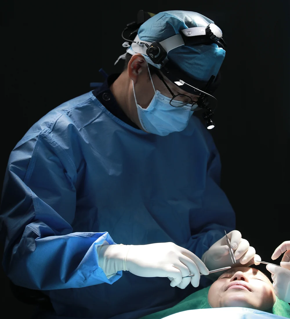
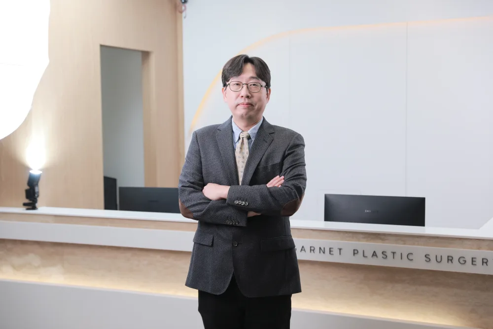
Lateral canthoplasty extends the outer corner of the eye, lengthening the visible eye opening and softening a steep outer angle. At Garnet it is planned and performed by one board-certified plastic surgeon, Dr. In-Soo Baek, from consultation through every follow-up.

A lateral canthoplasty is an eye-surgery procedure that extends the outer corner of the eye (the lateral canthus) to lengthen the visible palpebral fissure and reduce a steep outer slant. By repositioning the outer canthal angle - and, where planned, opening the lower-outer corner slightly - it is intended to give a longer, calmer eye shape that still suits your features.
The outer corner of the eye, where the upper and lower lids meet, sets much of the eye's length and slant. When the outer angle sits high or the eye looks horizontally short, the expression can read as sharper or smaller than a person wants. Unlike the inner corner, the outer corner has no single skin fold to release; the change comes from carefully repositioning the canthal angle itself.
Lateral canthoplasty extends this outer angle to lengthen the opening and soften the slant. Done conservatively it can also bring a touch of the lower-outer eye down, opening the eye shape. The technique matters: preserving a natural lateral canthal angle and avoiding over-correction are what keep the result looking unoperated and stable over time (Kim et al., Arch Plast Surg 2016; DOI 10.5999/aps.2016.43.4.316).
At Garnet this is a single-surgeon operation. Dr. Baek plans the case from the consultation, performs it himself, and reviews healing at set intervals; the clinic caps the day at two surgeries so each case has unhurried time. The stated aim is to address the concern you arrived with and to keep the outer corner looking like your own, rather than to chase the maximum possible opening.
From outer-corner assessment to the wide-angle extension and fine closure - every step by Dr. Baek.
A single surgeon, start to finish. Dr. Baek plans the case, performs the operation himself and reviews every follow-up. The clinic caps the day at two surgeries, so each operation has unhurried time.
Lateral canthoplasty typically takes about 30-60 minutes and is usually performed under local anaesthesia, with light sedation added where appropriate for comfort; the choice is decided with you and the anaesthesia team after your history is reviewed. The steps below outline how the wide-angle opening is carried out at Garnet.
Dr. Baek assesses the outer-corner angle, the eye length and slant, and the lower-lid support, and agrees the amount of extension and the design with you. Whether to combine inner-corner or lower-lid work is discussed here.
The extension is planned so the new outer corner sits in a natural position and the slant is softened, not flattened. The design is tailored to your eye shape and lower-lid tone so the result looks proportionate.
Working through a conjunctival incision at the outer corner, the canthal angle is extended; where planned, a small lower-canthal opening is added so the eye lengthens and opens without over-correcting. The canthal support is respected, not simply divided.
The outer corner is positioned and secured so it resists scarring back inward, and the lining and any skin are closed finely. Keeping the corner stable is what helps the lengthening hold over time.
Where the consultation shows it, an epicanthoplasty at the inner corner or a lower blepharoplasty is performed in the same sitting so the corners and the lid balance.
Dr. Baek reviews the closure himself. Because Garnet is single-surgeon, the doctor who planned and performed your case is the one who removes the sutures and follows you up.

Garnet's outer-corner technique is registered as the trademarked Wide-angle™ lateral canthoplasty, performed with an accompanying lower-canthal opening where indicated. Trademark registration describes the named technique, not a superior outcome.
At the outer corner, the upper and lower tarsal plates meet at the lateral canthal tendon, which anchors the lids to the bony orbital rim. Lengthening the eye here means working with this junction and the conjunctival lining rather than simply cutting skin, because the outer corner has a tendency to scar back toward its original position if the support is not handled carefully. Approaches that preserve or precisely reconstruct the canthal angle are associated with fewer complications and more stable results (Aesthet Surg J 2025; DOI 10.1093/asj/sjaf062).
Garnet's technique is a lateral canthoplasty with an accompanying lower-canthal (bottom) opening where indicated, planned for each eye so the outer corner is extended and the slant softened without over-opening. The same outer-corner principles inform the matching work sometimes combined with lower blepharoplasty, where the outer corner and the lower lid are considered together, and with round eye correction when the lid shape needs reshaping rather than simple lengthening.
| Lateral canthoplasty | Epicanthoplasty (medial) | Round-eye correction | |
|---|---|---|---|
| Corner addressed | Outer corner | Inner corner | Outer corner / lower lid |
| What it does | Extends the outer angle | Releases the inner fold | Reshapes a rounded or retracted lid |
| Effect on slant | Softens a steep slant | Little effect on slant | Restores a natural lid line |
| Common pairing | Lower blepharoplasty | Double-eyelid surgery | Canthoplasty / lid support |
| Typical use | Short or steep outer eye | Prominent inner fold | Over-rounded or scleral-show eye |
An integrated lateral canthoplasty that extends the fissure while preserving stability is described in Aesthet Surg J 2025 (DOI 10.1093/asj/sjaf062). The right corner - or combination - is individual; Dr. Baek advises at consultation, and the matching pages for epicanthoplasty and round eye correction explain each in detail.
Lateral canthoplasty is usually performed under local anaesthesia, with light sedation added where it helps comfort over a short procedure. Your medical history and any allergies are reviewed beforehand, and the plan is agreed with you and the anaesthesia team.
Because Garnet caps the day at two surgeries, the procedure is unhurried and the same surgeon who planned the case carries it out and reviews recovery - there is no separate operating doctor and no rotation of care.
Garnet is registered with Korea's foreign-patient programme; pre-operative checks, scheduling and after-care are coordinated for international visitors in English.
If an outer-corner extension would over-open the eye or risk reverting, or if a different combination suits you better, that is said at the consultation. Photos can be reviewed before you travel.
Much of a lateral canthoplasty is done through the conjunctiva (the inner lining) at the outer corner, so a significant part of the work leaves no external skin scar. Where a short skin extension is needed at the very outer corner, the line is placed in the natural outer-corner contour so it settles discreetly as it heals.
Any external mark is permanent but is designed to be inconspicuous once mature, and most are not obvious in everyday settings. The outer corner can be prone to scarring back inward, so stable fixation and a tension-aware closure matter; Dr. Baek reviews scar maturation and corner position at the 1-, 3- and 6-month visits and advises on care and, if ever needed, revision.
Outer-corner results are identifiable, so full before/after sets are reviewed privately at consultation with consent rather than published here. Results, recovery and suitability vary by individual and are not guaranteed.
Keep the head elevated early, use cold then warm compresses as advised, take medication as prescribed, keep the area clean and dry, and keep your follow-up visits.
Rubbing or pulling at the outer corners, eye make-up until cleared, strenuous exercise and bending early on, alcohol and smoking, and direct sun on the healing area.
Because lateral canthoplasty repositions the outer canthal angle structurally, the lengthening is intended to be a lasting change. Preserving the canthal angle and securing the corner stably are associated with results that hold and with fewer complications than over-aggressive techniques (Kim et al., Arch Plast Surg 2016; DOI 10.5999/aps.2016.43.4.316).
Individual results depend on the original anatomy, lower-lid support and how the corner heals. A known consideration is partial relapse, where the outer corner scars back toward its starting position; conservative, well-fixed planning lowers this risk, and any concern is assessed individually. The surrounding face continues to age normally, and the result does not prevent other eyelid procedures later.
Lateral canthoplasty is often planned with an epicanthoplasty at the inner corner, so both ends of the eye are balanced and the opening is lengthened evenly.
A lower blepharoplasty may be combined where under-eye bags or lower-lid skin are also a concern, and the outer corner and lower lid are treated as one unit.
Non-incision or incision double-eyelid surgery may be added where the upper-lid crease is part of the overall eye plan.
Where the lid shape - not just the length - needs reshaping, round eye correction may be planned alongside so the lid line and the corner are considered together.
Every operation carries risk. For lateral canthoplasty the most relevant concerns are partial relapse of the outer corner and, if extended too far or with weak lower-lid support, a pulled-down or rounded lower lid. Techniques that preserve the canthal angle and fix the corner stably are reported to reduce these complications (Arch Plast Surg 2016; DOI 10.5999/aps.2016.43.4.316). Careful patient selection is part of lowering the risk.
Other possible issues include temporary watering or irritation, mild asymmetry between the corners, conjunctival redness, over- or under-correction, and rarely a visible outer-corner scar. Over-opened outer corners can look unnatural, which is why Garnet plans a conservative extension. These are explained individually at consultation.
What reduces risk in practice: realistic planning of how far to extend, checking lower-lid support before opening, stable fixation, and follow-up by the operating surgeon. Garnet's single-surgeon, low-volume model is built around exactly this kind of unhurried planning and personal after-care.
Most international patients plan roughly 7-10 days in Korea for a lateral canthoplasty, so sutures can be removed by the surgeon before travel and the early swelling has settled. The coordinator confirms the timing for your specific plan, especially if you are combining it with inner-corner or lower-lid surgery.
Before you travel, send clear photos (front and a relaxed close-up of the outer corners) and a note on your concern and dates through WhatsApp, LINE or the form below. You'll get an honest pre-assessment - including whether an extension is worthwhile and stable for your anatomy - rather than a hard sell.
Garnet is registered with Korea's foreign-patient programme and coordinates consultations, scheduling and after-care in English. After you return home, Dr. Baek can continue to review your recovery by messenger.
| Lateral canthoplasty | Epicanthoplasty (medial) | Round-eye correction | |
|---|---|---|---|
| Corner addressed | Outer corner | Inner corner | Outer corner / lower lid |
| What it does | Extends the outer angle | Releases the inner fold | Reshapes a rounded or retracted lid |
| Effect on slant | Softens a steep slant | Little effect on slant | Restores a natural lid line |
| Common pairing | Lower blepharoplasty | Double-eyelid surgery | Canthoplasty / lid support |
| Typical use | Short or steep outer eye | Prominent inner fold | Over-rounded or scleral-show eye |
Citations are provided for general education. This page is informational and does not replace an in-person consultation; suitability, technique and recovery are individual.
Send photos and your question before you travel. An English-speaking coordinator reviews every enquiry and replies with honest guidance on whether surgery is appropriate, the likely plan and timing.
Prefer to chat now? Reach the coordinator directly: